Pheochromocytomas and paragangliomas represent rare but clinically significant neuroendocrine tumors that can cause serious health complications due to excessive catecholamine production. This article summarizes the latest findings in diagnostics, genetic testing, imaging methods as well as treatment options.
Pheochromocytomas (called intraadrenal paragangliomas) are among the rare neuroendocrine tumors that are located in the adrenal glands and produce catecholamines. Similar tumors that arise from tissue outside the adrenal glands, however, are called paragangliomas. Paragangliomas are most often found in the abdomen and less often in the pelvis, rarely in the chest (mediastinum) and in the neck and head.
From a clinical point of view, these tumors are mainly manifested by increased blood pressure (so-called hypertension) and accelerated heart rate (so-called tachycardia). Hypertension is present in about 90% of patients, half of whom have so-called paroxysmal hypertension. Seizures can last for several minutes or hours, often without cause, either day or night. In seizures, an accelerated heart rate is also often present (the patient often feels palpitations), pronounced sweating, pallor of the face, nervousness, anxious states, tremor. Seizures occur repeatedly, sometimes 2 times a day, sometimes 1 time per month or at other intervals. After an attack, the patient is often exhausted, tired, and sometimes may even have low blood pressure. If these tumors are not thought of, these seizures are often confused within the framework of various situations in which stress or even menopause plays a role.
The results of the latest genetic studies show that about 40% of these tumors are hereditary (about the same percentage is occupied by genetic abnormals/disorders that lead to tumor formation but are not hereditary). Therefore, today it is recommended that all patients with these tumors undergo a proper genetic examination, which includes not only consultation with a specialist in genetics, but also DNA tests from blood or saliva, which are used to detect gene mutations. Currently, 25 genes are known that are involved in the formation of these tumors, and according to the representation of certain genes on their formation, these tumors are divided into three basic groups.
The division of pheochromocytomas and paragangliomas into these three basic groups is of great clinical value, as the tumors of these groups differ greatly both in their clinical behavior and also in their detection using biochemical and imaging methods, and last but not least in their responses to different types of therapies.
From a clinical point of view, the most important are tumors that belong to the first group (cluster 1). These are mainly pheochromocytomas and paragangliomas, which arise from the mutation of succinate dehydrogenase (autosomal dominant inheritance) and often occur as early as childhood, have a great tendency to be multiple, recurrent and metastatic. Most of these tumors occur as paragangliomas outside the adrenal glands, primarily in the abdomen and in the head and neck region. The most aggressive are pheochromocytomas and paragangliomas, which arise on the basis of mutation of SDHB and SDHA. The most favorable clinical course is experienced by individuals with SDHC mutations.
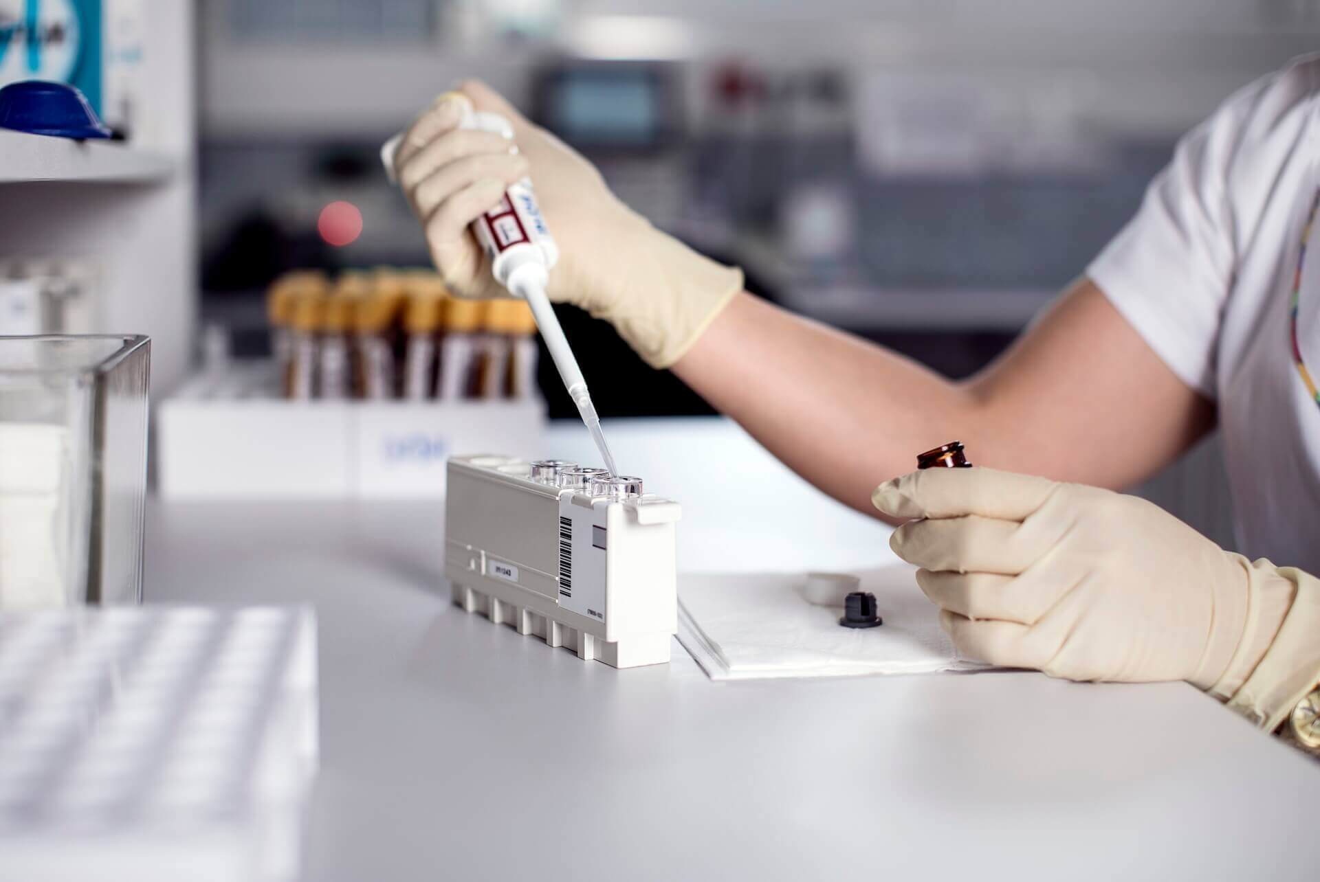
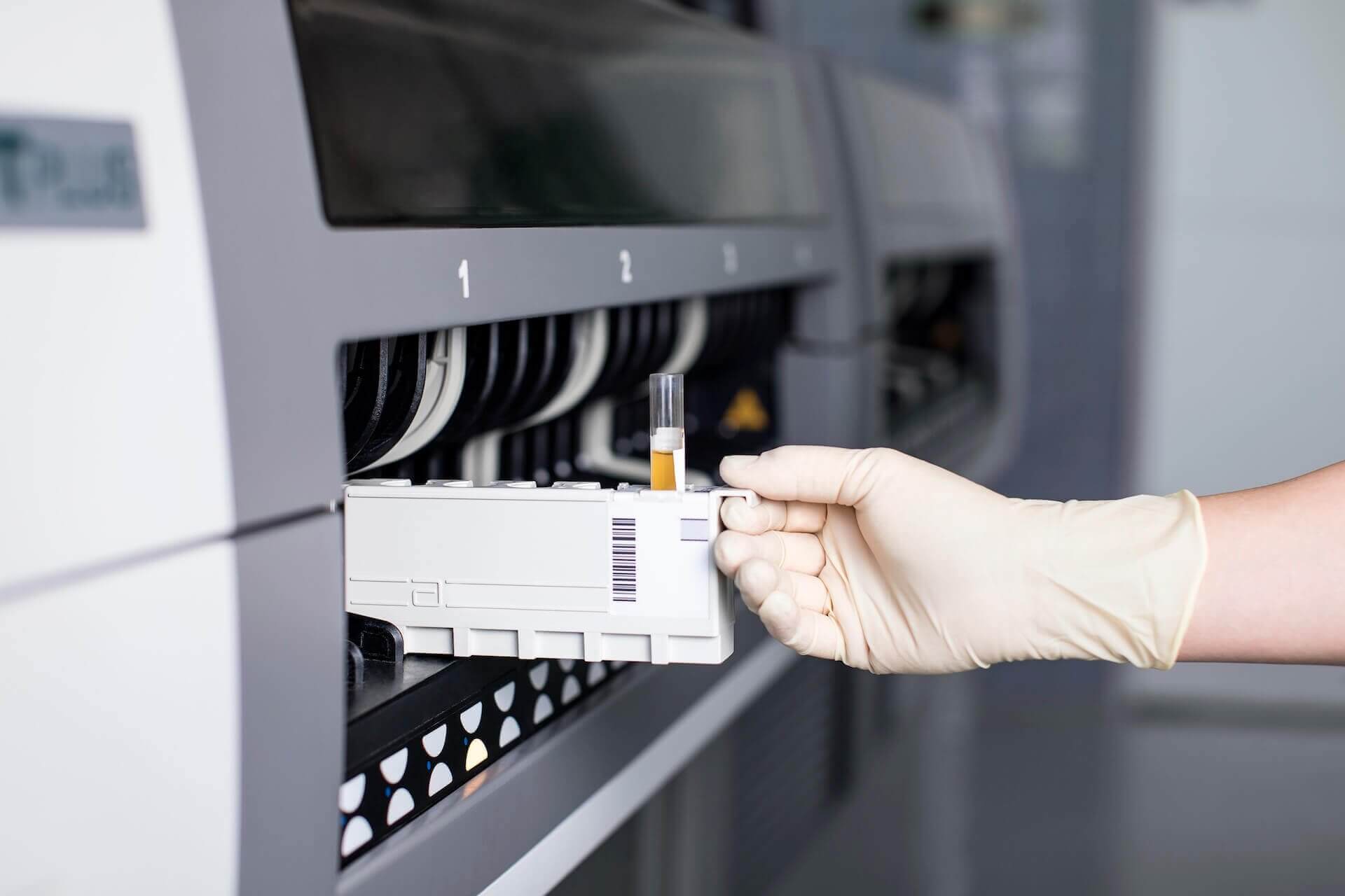
Currently, biochemical diagnosis of these tumors is based on the measurement of plasma or urinary free metanephrine. Due to the diagnostic capability of plasma metanephrine including easier blood collection than 24-hour urine collection, plasma metanephrine measurement is preferred today. Values that are 2 times higher than the upper limit of the norm indicate the presence of these tumors in most patients. In terms of biochemical diagnostics, the new biochemical marker 3-methoxythyramine is also measured, which is a reliable biomarker for the detection of pheochromocytomas and paragangliomas associated with gene mutations in the Krebs cycle.
In some patients, biochemical tests may be false positive due to antihypertensive or other medications. Sometimes a clonidine test needs to be performed in these patients. Biochemical examination must always be carried out lying down and at rest.
Every case is different. If you are not sure how to proceed further in a patient with a suspected adrenal tumor, please contact us. Together, we will assess the situation and recommend the next professional procedure — quickly, factually and in partnership.
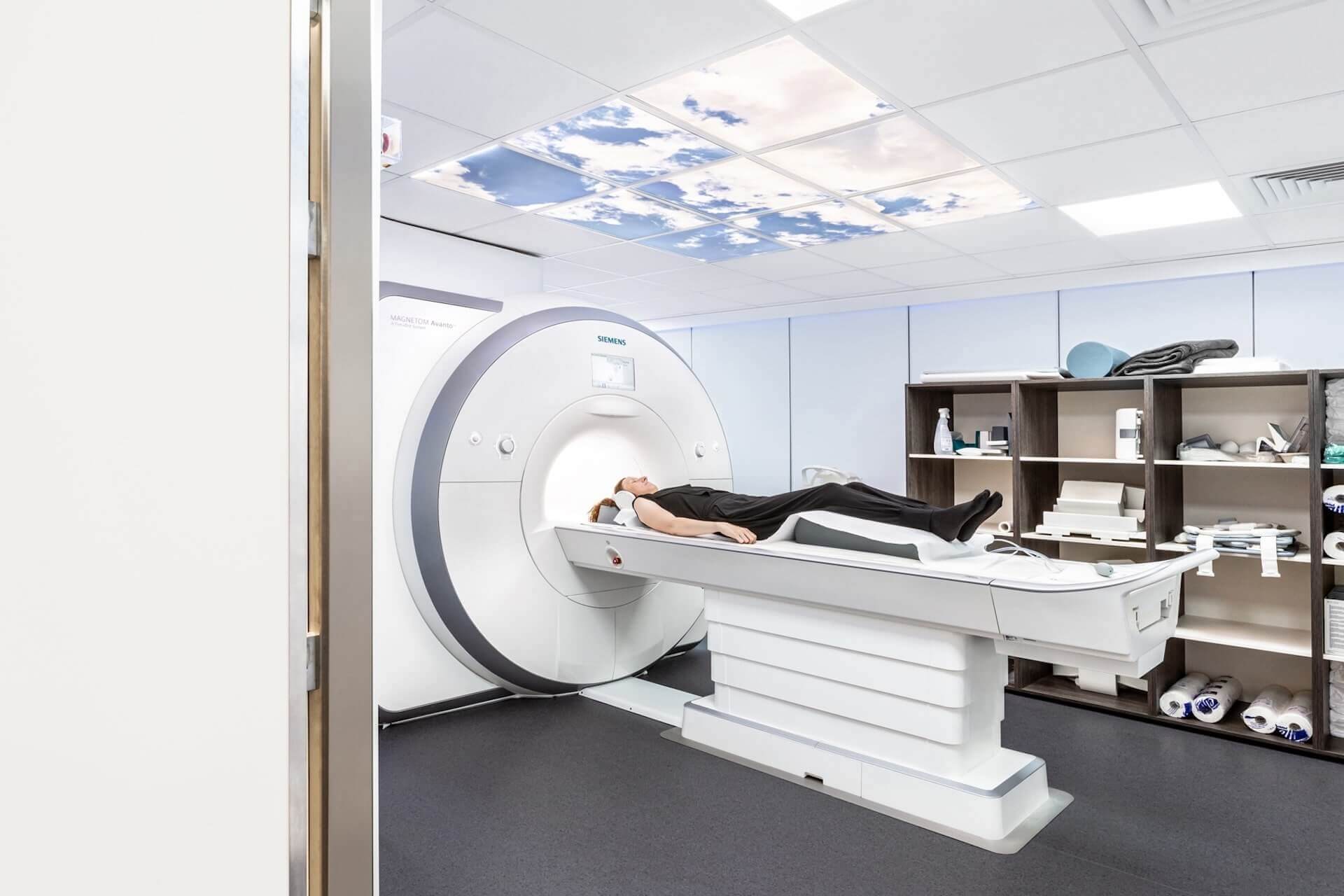
We approach the localization of pheochromocytomas and paragangliomas only if these tumors are really and correctly confirmed by biochemical examination. Tumors are first localized using anatomical imaging methods — CT or MRI. Since these tumors are very often multiple, metastatic or occur in places that may not be properly accessible to anatomical imaging methods, the so-called functional imaging methods are currently used (Department of Nuclear Medicine: so called. PET examination) inseparable from the correct and timely localization of these tumors.
With regard to these and other functional methods, 68Ga-DOTATATE and 68GA-DOTATOC, as well as FDG, 18F-FDOPA and 123I-MIBG are mainly used. It can also be noted that in terms of functional imaging methods, their importance also lies in the fact that, in the case of tumor positivity, these tumors are predisposed to possible radioactive treatment (see theranostic procedures) and these methods are also very suitable for regular monitoring of patients for possible exclusion of new multiple tumors, relapses and metastases.
Despite many new therapeutic procedures in the treatment of these tumors, the optimal therapy for pheochromocytomas and paragangliomas (excluding multiple metastases) is surgical removal of the tumor. Surgical solution should be approached for all pheochromocytomas and paragangliomas that are surgically accessible, even in very small tumors, as most of these tumors produce and release catecholamines that pose a serious threat to the patient's life if there is a sudden release of catecholamines from the tumor (e.g., during manipulation, psychological stress, necrosis) with a pronounced effect on the cardiovascular system (eg myocardial infarction, stroke, lethal arrhythmia, intestinal ischemia).
Metastatic tumors are treated with either chemotherapy (e.g., so-called chemotherapy). CVD, Temodar, etc.) or systemic radiotherapy (Lutathera, MIBG). Studies that focus on immunotherapy are at the beginning so far. Local radiotherapy is performed exceptionally in tumors that, for example, grow rapidly, often recur after surgery or significantly oppress surrounding tissues.
Since pheochromocytomas and paragangliomas represent very rare tumors, patients with these tumors should always seek very experienced specialists to diagnose and treat these tumors.

.jpg)